Slipped Capital Femoral Epiphysis
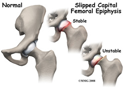
Image 35
Also known as proximal femoral epiphysiolysis, it is an alteration that occurs in adolescence and preadolescence. Slipped capital femoral epiphysis (SCFE) is the most common disorder of the hip in adolescents, especially during a period of rapid growth. SCFE presents most commonly between the ages of 11 to 15 years and is more common in male subjects. Obesity has been noted to be far more prevalent in patients with SCFE than in the normal population. Most commonly it affects patients with an adipose-genital phenotype, hormone imbalances, and other disorders in the presence of mechanical factors including excessive weight or trauma. It is typically idiopathic in nature although it can be associated with endocrinopathies and hypogonadism.
- Can be acute, acute-on-chronic, or chronic
- Clinically, the most common presenting symptom is hip pain. The pain is typically in the region of the groin but may be referred to the knee or anteromedial thigh. Thus it is important to consider SCFE in the adolescent athlete presenting with knee pain. The patient also may note a limp.
- If the slip is an acute one a dramatic onset of injury may be noted in the history with sudden severe hip pain and inability to bear weight or ambulate. The history of prodromal symptoms may be helpful in differentiating an acute SCFE from a true traumatic transphyseal fracture.
- The physical examination will reveal some pain and limitation of hip motion, especially internal rotation. Some have described the phenomenon of obligatory external rotation with flexion of the hip. In acute slips the leg may be held in external rotation and any attempt at range of motion may produce severe pain. In chronic slips there may be a mild leg length discrepancy and atrophy of the thigh.
Males > females and most common 10 -16 years of age. This condition affects African American children more frequently than Caucasian children. May be bilateral and present at different times. This is a fracture of the growth plate leading to a slipping of the femoral epiphysis off the femoral neck
Most common presentation is a varus slip
Related conditions
- Patient often overweight and there is an association with hypothyroidism.
- Patient complains of pain in hip or knee and holds the extremity externally rotated. Resists internal rotation and abduction: Not always linked to a specific incidence or mechanism of injury.
- Radiograph positive. The femoral head is displaced medially in relation to the femoral neck.
- Treatment is surgical with pinning of the joint
Most common presentation is a varus slip
- This is detected on a radiograph as a varus, posterior slipped proximal femoral epiphysis
- The “Klein line” is what is looked for on a radiograph
- The “Klein line” is a line that can be traced along the superior aspect of the femoral neck
- Failure to connect this line to the epiphysis is an early sign of slipped capital femoral epiphysis
- Slip tends to be progressive
- Valgus slips of the epiphysis are rare
Related conditions
- About 15% of acute slips result in avascular necrosis on presentation or following surgical pinning of the slipped capital femoral epiphysis.
- This condition can mimic a Salter I fracture with a slightly widened growth plate, if a frontal radiograph is the only view taken.
- In a child that has a chronic slip and continued pain, knowledge of the growth plate activity can help one to determine the necessity of continued immobilization or pinning.
- Slipped upper femoral epiphysis tends to occur in adolescents (aged 10-16 years), again more commonly in boys (3:1 ratio), patients of Afro-Caribbean origin, and obese patients
(5, 6, 7, 8, 20)
